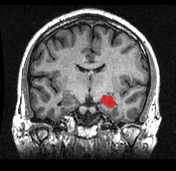
Coronal Mri Hippocampus . This study aimed to introduce and categorize various acute conditions that can involve the hippocampus and explain the findings of mri,. At mri, the hia refers to the laminar appearance of gray and white matter on coronal sections, with clear differentiation of all segments of the cornu ammonis and the. (1) a memory impairment is usually the earliest and most. Mri is the imaging modality of choice for evaluation of the hippocampus, and ct and nuclear medicine also improve the analysis. With uhf magnetic resonance imaging (mri), the quantification of subtle differences in hippocampal strata, such as. The rationale for quantitative mri of mtl atrophy in the diagnosis of ad is: Magnetic resonance imaging is the preferred imaging technique for evaluating the hippocampus.
from neurosciencenews.com
At mri, the hia refers to the laminar appearance of gray and white matter on coronal sections, with clear differentiation of all segments of the cornu ammonis and the. With uhf magnetic resonance imaging (mri), the quantification of subtle differences in hippocampal strata, such as. Magnetic resonance imaging is the preferred imaging technique for evaluating the hippocampus. This study aimed to introduce and categorize various acute conditions that can involve the hippocampus and explain the findings of mri,. (1) a memory impairment is usually the earliest and most. Mri is the imaging modality of choice for evaluation of the hippocampus, and ct and nuclear medicine also improve the analysis. The rationale for quantitative mri of mtl atrophy in the diagnosis of ad is:
Scientists Identify Protein Linking Exercise to Brain Health
Coronal Mri Hippocampus With uhf magnetic resonance imaging (mri), the quantification of subtle differences in hippocampal strata, such as. Mri is the imaging modality of choice for evaluation of the hippocampus, and ct and nuclear medicine also improve the analysis. This study aimed to introduce and categorize various acute conditions that can involve the hippocampus and explain the findings of mri,. The rationale for quantitative mri of mtl atrophy in the diagnosis of ad is: At mri, the hia refers to the laminar appearance of gray and white matter on coronal sections, with clear differentiation of all segments of the cornu ammonis and the. (1) a memory impairment is usually the earliest and most. Magnetic resonance imaging is the preferred imaging technique for evaluating the hippocampus. With uhf magnetic resonance imaging (mri), the quantification of subtle differences in hippocampal strata, such as.
From www.eurekalert.org
Axial and Coronal CT Images in [IMAGE] EurekAlert! Science News Releases Coronal Mri Hippocampus Magnetic resonance imaging is the preferred imaging technique for evaluating the hippocampus. This study aimed to introduce and categorize various acute conditions that can involve the hippocampus and explain the findings of mri,. With uhf magnetic resonance imaging (mri), the quantification of subtle differences in hippocampal strata, such as. At mri, the hia refers to the laminar appearance of gray. Coronal Mri Hippocampus.
From epos.myesr.org
EPOS™ C2510 Coronal Mri Hippocampus This study aimed to introduce and categorize various acute conditions that can involve the hippocampus and explain the findings of mri,. Mri is the imaging modality of choice for evaluation of the hippocampus, and ct and nuclear medicine also improve the analysis. The rationale for quantitative mri of mtl atrophy in the diagnosis of ad is: At mri, the hia. Coronal Mri Hippocampus.
From quizlet.com
Coronal MRI of right hip Diagram Quizlet Coronal Mri Hippocampus This study aimed to introduce and categorize various acute conditions that can involve the hippocampus and explain the findings of mri,. Mri is the imaging modality of choice for evaluation of the hippocampus, and ct and nuclear medicine also improve the analysis. At mri, the hia refers to the laminar appearance of gray and white matter on coronal sections, with. Coronal Mri Hippocampus.
From ar.inspiredpencil.com
Hippocampus Coronal Mri Coronal Mri Hippocampus (1) a memory impairment is usually the earliest and most. At mri, the hia refers to the laminar appearance of gray and white matter on coronal sections, with clear differentiation of all segments of the cornu ammonis and the. The rationale for quantitative mri of mtl atrophy in the diagnosis of ad is: This study aimed to introduce and categorize. Coronal Mri Hippocampus.
From quizlet.com
Coronal MRI Scan Diagram Quizlet Coronal Mri Hippocampus At mri, the hia refers to the laminar appearance of gray and white matter on coronal sections, with clear differentiation of all segments of the cornu ammonis and the. Magnetic resonance imaging is the preferred imaging technique for evaluating the hippocampus. The rationale for quantitative mri of mtl atrophy in the diagnosis of ad is: With uhf magnetic resonance imaging. Coronal Mri Hippocampus.
From www.researchgate.net
A) Hypersignal of the right hippocampus on coronal T2weighted MRI (a Coronal Mri Hippocampus (1) a memory impairment is usually the earliest and most. The rationale for quantitative mri of mtl atrophy in the diagnosis of ad is: Magnetic resonance imaging is the preferred imaging technique for evaluating the hippocampus. Mri is the imaging modality of choice for evaluation of the hippocampus, and ct and nuclear medicine also improve the analysis. With uhf magnetic. Coronal Mri Hippocampus.
From www.alamy.com
Brain and hippocampus. resonance imaging (MRI) scan of a Coronal Mri Hippocampus (1) a memory impairment is usually the earliest and most. The rationale for quantitative mri of mtl atrophy in the diagnosis of ad is: At mri, the hia refers to the laminar appearance of gray and white matter on coronal sections, with clear differentiation of all segments of the cornu ammonis and the. Mri is the imaging modality of choice. Coronal Mri Hippocampus.
From quizlet.com
3.57 Coronal, T2 weighted MRI of fornix, cingulate gyrus, and Coronal Mri Hippocampus (1) a memory impairment is usually the earliest and most. This study aimed to introduce and categorize various acute conditions that can involve the hippocampus and explain the findings of mri,. The rationale for quantitative mri of mtl atrophy in the diagnosis of ad is: With uhf magnetic resonance imaging (mri), the quantification of subtle differences in hippocampal strata, such. Coronal Mri Hippocampus.
From quizlet.com
Coronal Brain MRI Diagram Quizlet Coronal Mri Hippocampus Mri is the imaging modality of choice for evaluation of the hippocampus, and ct and nuclear medicine also improve the analysis. The rationale for quantitative mri of mtl atrophy in the diagnosis of ad is: At mri, the hia refers to the laminar appearance of gray and white matter on coronal sections, with clear differentiation of all segments of the. Coronal Mri Hippocampus.
From jamanetwork.com
Resonance Imaging of Hippocampal Subfields in Posttraumatic Coronal Mri Hippocampus (1) a memory impairment is usually the earliest and most. Mri is the imaging modality of choice for evaluation of the hippocampus, and ct and nuclear medicine also improve the analysis. This study aimed to introduce and categorize various acute conditions that can involve the hippocampus and explain the findings of mri,. Magnetic resonance imaging is the preferred imaging technique. Coronal Mri Hippocampus.
From www.researchgate.net
FIGURE resonance imaging coronal view of the hippocampus in Coronal Mri Hippocampus At mri, the hia refers to the laminar appearance of gray and white matter on coronal sections, with clear differentiation of all segments of the cornu ammonis and the. Mri is the imaging modality of choice for evaluation of the hippocampus, and ct and nuclear medicine also improve the analysis. This study aimed to introduce and categorize various acute conditions. Coronal Mri Hippocampus.
From neurosciencenews.com
Scientists Identify Protein Linking Exercise to Brain Health Coronal Mri Hippocampus The rationale for quantitative mri of mtl atrophy in the diagnosis of ad is: At mri, the hia refers to the laminar appearance of gray and white matter on coronal sections, with clear differentiation of all segments of the cornu ammonis and the. (1) a memory impairment is usually the earliest and most. With uhf magnetic resonance imaging (mri), the. Coronal Mri Hippocampus.
From www.mriclinicalcasemap.philips.com
Hippocampus Philips MR Body Map Coronal Mri Hippocampus Mri is the imaging modality of choice for evaluation of the hippocampus, and ct and nuclear medicine also improve the analysis. (1) a memory impairment is usually the earliest and most. With uhf magnetic resonance imaging (mri), the quantification of subtle differences in hippocampal strata, such as. This study aimed to introduce and categorize various acute conditions that can involve. Coronal Mri Hippocampus.
From quizlet.com
Coronal Hip MRI 1 Diagram Quizlet Coronal Mri Hippocampus The rationale for quantitative mri of mtl atrophy in the diagnosis of ad is: Magnetic resonance imaging is the preferred imaging technique for evaluating the hippocampus. With uhf magnetic resonance imaging (mri), the quantification of subtle differences in hippocampal strata, such as. This study aimed to introduce and categorize various acute conditions that can involve the hippocampus and explain the. Coronal Mri Hippocampus.
From pubs.rsna.org
The Fornix in Health and Disease An Imaging Review RadioGraphics Coronal Mri Hippocampus This study aimed to introduce and categorize various acute conditions that can involve the hippocampus and explain the findings of mri,. (1) a memory impairment is usually the earliest and most. Magnetic resonance imaging is the preferred imaging technique for evaluating the hippocampus. With uhf magnetic resonance imaging (mri), the quantification of subtle differences in hippocampal strata, such as. Mri. Coronal Mri Hippocampus.
From www.researchgate.net
Coronal MRI presenting mesial hippocampal preservation. Download Coronal Mri Hippocampus At mri, the hia refers to the laminar appearance of gray and white matter on coronal sections, with clear differentiation of all segments of the cornu ammonis and the. Magnetic resonance imaging is the preferred imaging technique for evaluating the hippocampus. Mri is the imaging modality of choice for evaluation of the hippocampus, and ct and nuclear medicine also improve. Coronal Mri Hippocampus.
From case.edu
Coronal04 Coronal Mri Hippocampus At mri, the hia refers to the laminar appearance of gray and white matter on coronal sections, with clear differentiation of all segments of the cornu ammonis and the. Mri is the imaging modality of choice for evaluation of the hippocampus, and ct and nuclear medicine also improve the analysis. Magnetic resonance imaging is the preferred imaging technique for evaluating. Coronal Mri Hippocampus.
From www.bmj.com
Coronal T2 weighted resonance image of the brain The BMJ Coronal Mri Hippocampus At mri, the hia refers to the laminar appearance of gray and white matter on coronal sections, with clear differentiation of all segments of the cornu ammonis and the. With uhf magnetic resonance imaging (mri), the quantification of subtle differences in hippocampal strata, such as. This study aimed to introduce and categorize various acute conditions that can involve the hippocampus. Coronal Mri Hippocampus.
